|
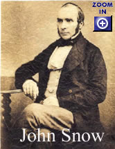
Cholera spread rapidly throughout the world after the 1817 epidemic, largely due to the inadvertent transport of bilge water, mainly from British ships, but others too, acquired in the Bay of Bengal that contained the organisms. Dumping the contaminated water into their own port cites upon arrival home seeded the local waters with it and insured the eventuality of an outbreak. It then rapidly moved throughout Europe and into Russia. The French were the ones who brought it to the New World, and in 1832, it spread south from Montreal and caused an enormous epidemicin New York City (see: The Cholera Years, by C. E. Rosenberg, The University of Chicago Press, 1987, pp 265). In 1855, a wave of cholera ravaged the citizens of some parts of London. Thousands became ill and died before the medical detective work by John Snow identified the Broad Street water pump as the single point source of that outbreak. His classical maps showing where people who became sick lived convinced him that the only possible source of the infection was the water pump. This landmark study established the epidemiological view of cholera that has endured until quite recently. In London on the corner of Broadwick (formerly Broad) stands the John Snow pub, a fitting commemorative honoring the site on which these historic events unfolded. Today, all patrons of the John Snow can enjoy a pint of local ale, and even more importantly, a refreshing glass of crystal clear, pathogen-free water. Since 1961, there have been seven major cholera pandemics (The global spread of cholera during the seventh pandemic, 1961-1971 [source]), affectingmillions of people living in South America, Africa, Europe, and Asia. To fully appreciate its biology, one must take into account data collected from many different scientific disciplines. Ecology, molecular biology, microbiology, epidemiology, pathology, and long range sensing all have supplied critical pieces of information, which, taken together and integrated, forms a comprehensive body of knowledge as to how cholera enters the human population and what factors regulate its occurrence within the estuary. Thus, cholera is a perfectly suited topic for illustrating the usefulness of the Medical Ecology paradigm. The Cholera Organism
First isolated, cultured, and characterized by Robert Koch in Germany in 1883, the organism is a comma-shaped, flagellated, gram-negative bacterium, Vibrio cholerae. In fact, it was Koch’s work on cholera the led the way to firmly establishing the germ theory of disease, and helped convince the medical community as to the microbial nature of this devastating clinical condition. For all of his exemplary work, he was the recipient of the Nobel Prize in Medicine in 1905. In the laboratory, it can be easily grown at 37°C on blood agar, as well as on selective media such as thio-citrate-bile salt-sucrose. There are many 16 strains of V. cholerae, and the 01 and 0139 strains are the most lethal. While V. cholerae is the best characterized of these agents, several other species of Vibrio can also cause significant disease. Pathological strains produce clinical symptoms and signs, the most common one by far being a protracted, watery diarrhea. Yet, despite the fact that human populations are routinely infected with it, V. cholerae’s natural habitat is not our small intestine, since most infections last for only several days, and the carrier state in humans is extremely rare. It was well into the 21st century before its fundamental niche was revealed to be the estuary, a narrow ecological region known as an ecotone. Typically, the first clinical cases of any new outbreak occur in communities situated on or near an estuary. Although this fact was known for at least since the 1800s, it was not considered essential to the natural history of the disease. In fact, its ecological role, once revealed, surprised even those who resolutely suspected that it was essentially an organism that occupied a fundamental niche outside the human host, but could not prove it. How Do Cholera Epidemics Start?One of the enigmas related both to its ecology and to human disease was its apparent absence from human populations just prior to epidemics. Besides the fact that extensive clinical research repeatedly failed to identify human carrier states as the source, the bacterium did not form spores, so a resting stage could not be demonstrated in the estuarian environment. V. cholerae seemed to simply “disappear” at times when cases were not occurring (prior to the arrival of the monsoons in South Asia, for example). How does an epidemic get its start if humans are not the source of the initial infection? Could there be reservoir animal species that harbored the organism and occasionally contaminated the human environment, or perhaps there was a stage of the bacterium that was more difficult to find that a spore stage that allowed it to survive in salt water. Monsoons represent seasonal patterns of precipitation that bring with them changes in both the relative salinity and temperature of the estuaries of major river systems along the entire coast of the Indian subcontinent. These and other related seasonal precipitation events in similar tropical and sub-tropical environments cause dramatic shifts in the aquatic environment, triggering blooms of phytoplankton, that in turn serve as the food source for a rich assortment of zooplankton grazer species. It has been known from the time of Koch that cholera organisms grow best at a temperatures above 17°C, and in a nutrient broth with a [NaCl] of 5-15 parts per million, well below that of the open ocean, but above that of freshwater. Those conditions are met in the estuaries by episodes of heavy rains in the spring, and appear to be absolutely essential to establishing an ecological setting favoring an outbreak. But where do the organisms come from? After years of intensive laboratory and field studies, it was discovered that many species of copepods that comprise the myriad assemblages of zooplankton communities in those estuaries harbor V. cholerae as an ecto-symbiont. Organisms can be found growing on their egg sacks and inside their gut tracts. This discovery opened the way for a more complete description of the ecology of the cholera group of bacteria. There still, however, remained unanswered questions regarding its epidemic nature. One large missing piece of the puzzle was the fact that zooplankton blooms are not always present in the estuary, and hence cholera cannot routinely be cultured from most brackish water environments. Where did the microorganisms go during quiescent periods in between seasonal rain events? Further laboratory-based research revealed that they could transform into a unique dormant stage that was able to survive for months in the sediment of the estuary. This stage was unlike spores of bacteria such as Bacillus subtilis, an aerobic, non-pathogenic, soil-dwelling organism, or its anaerobic cousin, Clostridium perfringens, a soil-dwelling bacterium capable of causing acute gastroenteritis and gas gangrene. These exciting new data enabled investigators to now integrate information regarding the seasonal nature of events surrounding an outbreak in populations living near estuaries with previous data on the physical and chemical requirements for its growth. What has emerged over the last 10 years has quite literally revolutionized the way we look at cholera. A Modern SynthesisSeasonally dependent epidemics can now be described in ecological terms. Warm, intense episodes of precipitation falling on coastal and nearby inland regions transforms the temperature and salinity profiles of estuaries. These changes create favorable growth conditions for the dormant bacterium, and also, most importantly, for phytoplankton species. The influx of large quantities of freshwater mobilizes stored nutrients in the bottom sediments of the estuary, and gives the cholera bacterium a head start in its growth cycle. Algal blooms in response to higher temperatures, lower salinity, and nutrient loading allow for a similar increase in filter feeding zooplankton. Nutrient loading of the estuary typically occurs from a variety of nearby riparian sites (point source and non-point source run off), thus serving as the final environmental cue, supplying V. cholerae with additional sources of nutrients. This apparently is sufficient enough to permit an increase in bacterial cell numbers to a level that enables the organism to encounter copepods of the right species. Copepod species then pick up the bacteria on their external and internal surfaces. This most likely involves specific bacterial ligands and copepod surface receptor molecules, yet to be identified and characterized. Once the cholera organism attaches, the crustaceans carry the bacteria along as a normal component of their bodies throughout their life cycle. The bacterium continues to replicate until they completely cover the surface of the copepod’s egg sack. When eggs mature, the overall process apparently triggers the cholera bacteria to synthesize, then secrete a chitinase that functions to dissolve the outer egg case, facilitating the release of the eggs, dispersing them and the bacteria into the water column. The more dense the population of zooplankton, the more the concentration of free cholera bacteria there will be in the water column. Filter feeding benthic organisms (e.g. crabs, clams, and oysters) process large volumes of water, concentrating particulates and the cholera bacteria in their gut tracts. Humans that harvest these contaminated food organisms in spring-time, and ingest them raw place themselves and the rest of their local communities at risk from acquiring cholera. A bacterial cell concentration of 103/ml of water is necessary to allow an infectious dose of V. cholerae to accumulate within mollusks and crustaceans. Application of Ecological Knowledge to the Control of Cholera
|
|||||||||||||||||||||||||||||||||||||||||||||


|